
The function of the eyes is to receive and guide light energy (photons) to specialized receptor cells (photoreceptors) that translate this photonic energy into electrical energy. Then transmitted to the brain by neurons, it will be analyzed and interpreted to give birth to a mental representation of the environment allowing the animal to move; to locate its prey or its sexual partner, etc.
Vertebrate eyes, made of a single structure, are called simple eyes. In arthropods, the eyes are called composites because they consist of several similar subunits ommatidies .
Compound eyes appeared early in evolution. They have been observed on Cambrian crustacean and chelerate fossils (scorpions and spiders) (540-485 million years) and Devonian insects (420-360 Ma). This period was favourable to a great diversification of the animal world. The eyes composed of the current horseshoe crab (Limulus sp., Chelerates) have thus remained unchanged since that time (read this article).
Because of their small size, the observation and study of insect eyes had to wait for the invention of the microscope in the seventeenth century. The first descriptions were made by the English chemist, physicist and mathematician Robert Hooke in 1665, by the Dutch scientist Antoni Van Leeuwenhoek in 1695 and by the Austrian physiologist Sigmund Exner in 1891.
I) Presence of compound eyes
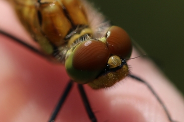
The compound eyes are found in almost all insect species
The compound eyes are found in almost all insect species
The compound eyes are found in almost all insect species
The compound eyes are found in almost all insect species
“text-align=”>”text-align=”>.
In the Aptérygotes (wingless), insects qualified as “primitive”, they are absent for example in Thysanurae (order of apterous insects) and Proturae (arthropods pancrustacés hexapodes long considered as insects) and consist only of 8 ommatidies in others.
By contrast, among Ptérygotes (winged), insects qualified as “modern”, the number of ommatidies can be particularly high : 800 among Drosophila (Diptera), 7 500 among Diopsidae (Diptera) (read this article), 10 000 in bumblebees (Hymenoptera), or 30 000 in Odonates (dragonflies).
Some cavernicolous species, underground like termites (Isoptera), parasites with atrophied wings (Phthiraptera and Siphonaptera), or mealybugs (Hemiptera) are devoid of them. The eye of the ant Hyponera punctatissima (Hymenorptera – Formicidae) consists of only one ommatidie.
II) Structure of an ommatidie
Each ommatidie consists of 3 parts : optical, light collection and sensory.
Alike the whole body of insects, compound eyes are covered with a cuticle (read this article), but for obvious reasons, this one is in this transparent place, colourless and forms a cornea, or biconvex lens, on the surface of each ommatidie.
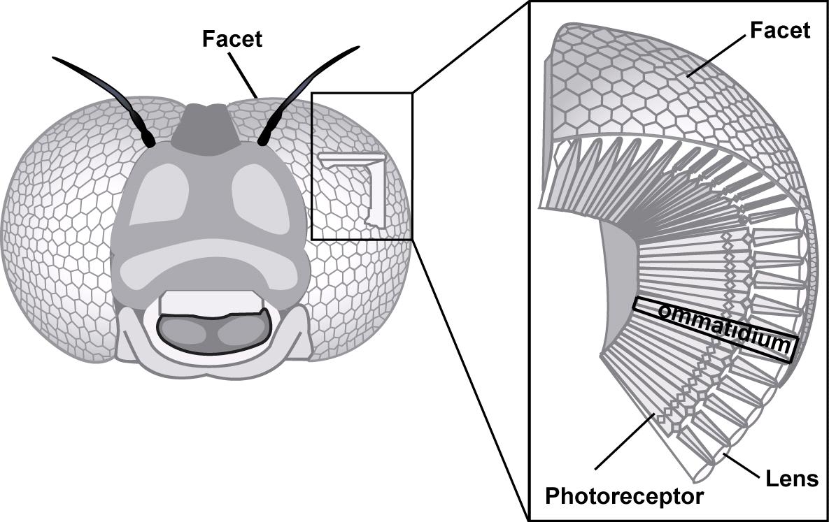
View from above, ommatidies form hexagonal facets placed next to each other (figure 1).
The cornea is synthesized and secreted by two epidermal cells, corneal cells, or also primary pigment cells. Under the cornea are 4 cells (cells Semper) whose function is to produce, in certain species, a second rigid and transparent lens called crystalline cone (Figure 2). Under the cornea and the crystalline cone are placed the sensory cells (photoreceptors or retinal cells) which are elongated neurons, generally 8, sometimes 7 or 9, laterally bordered by 12 to 18 supporting cells, separating the ommatidies between them, and called secondary pigmentary cells (figure 2). Photoreceptors pass through the basal lamina where they connect to the optic nerve neurons at the optic lobe.
The internal margin of photoreceptors is composed of very dense microvilli and perpendicular to the axis of the cell rhabdomere, where visual pigments like rhodopsin.
are located caption id=”attachment_2591″ align=”alignleft” width=”251″] Figure 2 : General structure of an insect ommatidie (Source : The Insects : Structure and Function – R.F. Chapman – 5th Edition (2013) – p710 – Modified by B. GILLES)[/caption]

The union of the microvilli of all photoreceptors forms the rhabdome ( see figure 3).
In some species of bees or flies, the photoreceptors twist along their length thus modifying the orientation of the microvillosity, which has the effect of eliminating the effects of polarization (explanation in a future article).
Rhabdomeres can be either “merged” or “open”. In the first case, all rhabdomes are in contact with each other and have the same field of vision. In the second, they are separated by a matrix and each have their own visual field (see figure 4).
caption id=”attachment_2604″ align=”aligncenter” width=”679″] Figure 4 : Rhabdomères “fusionnés” (left) – Rhabdomères “ouverts” (right) (Source : The Insects : Structure and Function – R.F. Chapman – 5th Edition (2013) – p710 and 712 – Modified by B. GILLES)[/caption]
The configuration of compound eyes differs if the insects are day or night. The amount of light being lower at night, evolution has modified the structure of ommatidies and compound eyes to increase photon capture efficiency.
- Structure in apposition : it occurs mainly in daytime species. The rhabdome extends over the entire length of the photoreceptor cell, from the crystalline cone to the basal blade, i.e. a length of 150 µm in ants Camponotus and 80 µm in Drosophila flies ( see figure 5A). Photoreceptors receive light only from the lens to which they are attached.
- Structure in superposition : it occurs mainly in nocturnal species (butterflies and beetles). The rhabdome does not extend over the entire length of the photoreceptor cell ( see figure 5B). Photoreceptors receive light from hundreds, even thousands, of facets increasing photon capture and thus visual sensitivity.
caption id=”attachment_2606″ align=”aligncenter” width=”598″] Figure 5 : A) Cross-section of an eye composed of apposition type (day insects) – B) Cross-section of an eye composed of superposition type (nocturnal insects) (Source : Warrant, 2008 – Modified by B. GILLES)[/caption]
Notes: Hymenoptera (wasps, bees, ants) have eyes of type apposition. Although this type of vision offers low light sensitivity, some species, which became nocturnal during evolution, have good visual acuity against all odds. Many scientists, like the Australian Eric J. Warrant (2008), are conducting experiments to unravel this mystery.
12 to 18 secondary pigment cells isolate ommatidies from each other.
Another adaptation to night life consists, especially in butterflies, of flattening and widening the respiratory trachea at the back of the eye to form a layer called tapetum. The function of this structure is to reflect light. Photons pass through rhabdomes twice, increasing the number of excited visual pigments and thus the sensitivity of vision.
A video describing all this as an animation
III) External adaptation of ommatidies
The form and arrangement of ommatidies differ between insect species.
For example, in Mantopters, the capture of prey requires high resolution binocular vision. For that, evolution has endowed them with compound eyes where the diameter of the facets, the interommatidal angle and the length of the rhabdomes of the ommatidies are more important in the center to decrease towards the peripheries ( see figure 6). These characteristics offer to the insect a visual zone of great precision called fovéa (see infography below). These adaptations are also found in the eyes of vertebrates.
caption id=”attachment_2610″ align=”aligncenter” width=”858″] Figure 6 : Variation of ommatidies in Tenodera (Mantoptera) from Rossel, 1979 – Frontal view of the head (left) – Compound eye with open fovea (middle) – Cross section of the compound eye showing differences in curvature, interommatidal angle and rhabdome length (right) (Source : The Insects : Structure and Function – R.F. Chapman – 5th Edition (2013) – p714 – Modified by B. GILLES)[/caption]caption id=”attachment_3421″ align=”alignleft” width=”160″]
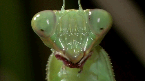 Tête de mante-religieuse – the fovea are the black spots in the middle of the compound eyes (Source : media.giphy)[/caption]
The area of high visual resolution is located in front of the compound eyes in the mantises-religious (infography opposite), on the fronto-dorsal part of the eyes composed of dragonflies (Odonates) which hunt and pursue aerial prey. Other insects, such as Beetles of the genus Gyrinus (family Gyrinidae) which swim on the surface of the water, have two areas of high visual resolution, one below the surface (ventral) and the second above (dorsal). Even more surprising, the eyes composed of males of the genus Cloeon (Ephemeroptera) are made up of two independent parts. One, dorsal, is made up of ommatidies with large facets and of superposition type and the other, lateral, with smaller facets and of affosition type (see photo album below).
IV) Internal adaptation of ommatidies
The shape of the lens varies between insect species. It is generally conical, except for so-called “primitive” insects such as Collembola and Thysanurae in which it is spherical.
It has been described 4 types of crystalline cone
- Eucône : Semper cells are located in the crystalline cone (figure 7)
- Acône : absence of crystalline cone, the role of the focus is played by Semper cells whose cytoplasm is clear. This pattern occurs in Coccinellidae, Staphylinidae, Tenebrionidae, some Diptera and Hemiptera
- Pseudocône : Semper cells secrete an extracellular crystalline cone formed of gelatinous liquid (some Odonates, Coleoptera and most Diptera)
- Exocône : Semper cells do not contribute to the development, it is the contact lens which, by forming a cone, plays this role (Elateridae and Lampyridae) (Figure 8)
Photoreceptors are generally of the same length. However, in so-called “primitive” insect species such as the silverfish (Thysanurae) and butterflies of the genus Spodoptera, the photoreceptors are wound and arranged in a stepped manner. Other configurations are also possible (figure 9).
caption id=”attachment_2614″ align=”alignright” width=”250″] Figure 8 : Exocone-like Ommatidie whose clear zone is formed by Semper cells – Adaptation to night (left) – Adaptation to day (right) (In Photuris, Coleoptera species) – From Walcott, 1975 (Source : The Insects : Structure and Function – R.F. Chapman – 5th Edition (2013)- p711 – Modified by B. GILLES)[/caption]


For those who wish to continue, find the second article which is devoted to the perception of shapes, distances and speed. Follow this link.
The functioning of all these structures and the processing of visual information will be the subject of future articles (to be informed, subscribe to the newsletter if it is not yet done !)
Source :
– The Insects : Structure & Function (5th edition, 2013) – Edition : S.J. Simpson & A.E. Douglas – Cambridge University Press
– Encyclopedia of Insects : Vincent H. RESH & Ring T. CARDE, Academic Press-2009
– Srinivasan M. V. (2011) : Honeybees as a model for the study of visually guided flight, navigation and biologically inspired robotics – American Physiological Society, Vol.91 (2) 413-460 (lien)
– Warrant E. J. (2008) : Seeing in the dark : vision and visual behavior in nocturnal bees and wasps – Journal of Experimental Biology, 211:1737-1746 (lien)
Recommendation of books on this theme
>
– Evolution of the Insects (David Grimaldi & Michael S. Engel – Edition : Cambridge University Press – 772 pages – May 16, 2005)
– The Insects: An Outline of Entomology (P.J. Gullan & P.S. Cranston – Edition : Wiley-Blackwell – 624 pages – 5ème édition : 6 août 2014)
– The Insects: Structure and Function (R.F. Chapman & Stephen J. Simpson – Edition : Cambridge University Press – 959 pages – 5ème édition : 12 novembre 2012)
– Encyclopedia of Insects (Vincent H. Resh & Ring T. Cardé – Edition : Academic Press – 1024 pages – 2ème édition : 22 juillet 2009)