Sensory receptors in insects: mechanoreceptors – Part 3: tension and stretch receptors
Third part : tension and stretch receptors (prioceptors)
Alike all arthropods, insects have an external skeleton, the exoskeleton, consisting of chitin and forming the cuticle (lire th article). Its insulating properties imply that the cuticle creates a barrier to the reception of stimuli from the external environment to the internal physiological environment. In order to circumvent this problem, evolution has given rise to a whole range of structures designed to transmit a wide range of types of stimuli to the insect, enabling it to grasp its environment effectively and accurately and to respond favourably to any modification by an adapted behaviour.
Among these sensory structures, some are sensitive to mechanical distortions (mechanoreceptors object of this article), others to light (photoreceptors) and others to chemical components (checeptors or chemocheceptors).
I) General
Mechanoreceptors are classified into three main categories (Table I)
| Categories | Anatomical position | Types | Functions |
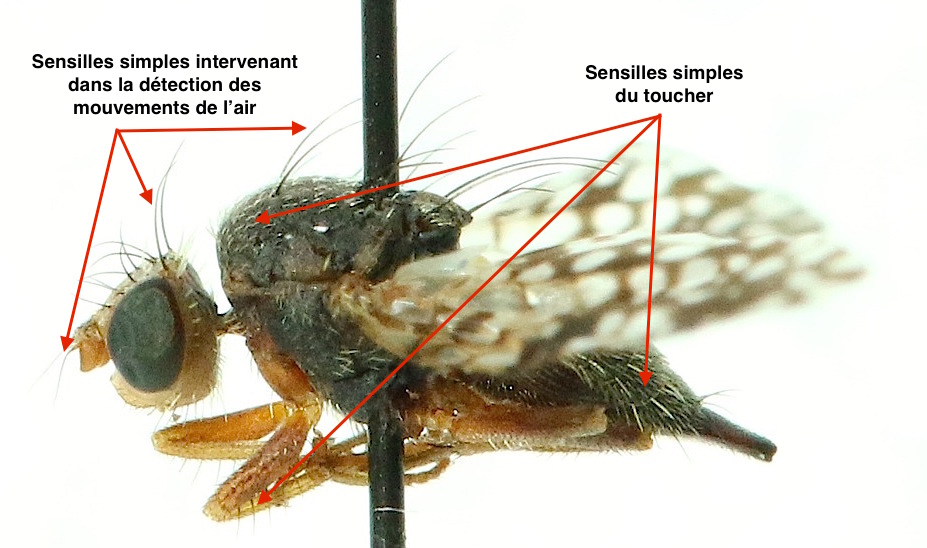
| Extérorécepteurs Cuticular receivers (Click here) | Cuticle surface | Senseilles simples | Touch, pressure variation (air or water), gravity detection, proprioception* |
| Campaniforms | Cup pressure variation | ||
| Interoreceptors Intradermal or subcuticular receptors (click here) | Derm | Chordotonal organs | Pressure variation (air, water), gravity detection, proprioception* |
| Property receivers | Internal fabrics | No precise structure | Proprioception* |
| * Proprioception : perception of the position of the different parts of the body | |||
Featuring amazing and multiple properties, they can intervene in the sense of touch when stimulated by contact with an object or substrate, in the sense of hearing by stimulation of air or water and also in the control of posture and movement when excited by movements of parts of the insect’s body.
II) Structural description and function
>
Tension and stretch receptors (also called « stretchable ») are simple structures, unlike other types of mechanoreceptors: cuticular (lire part 1) and chordotonal (lire part 2).
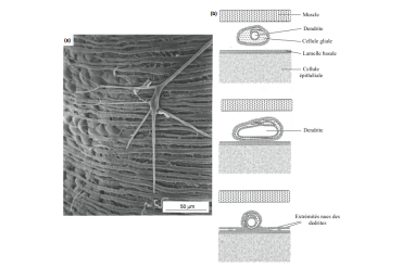
caption id= »attachment_2389″ align= »alignright » width= »360″] Illustration 1 : Non-specialized stretching receptors (after Sugawara, 1981) – a) Free multipolar neuron in the hemolymph where dendritic endings pass between the epithelium and muscle fibers – b) cross sections of dendrite below a muscle layer (Source : The Insects : Structure and Function – R.F. Chapman – 5th Edition (2013) -p765 – Modified by Benoît GILLES)[/caption]
Their structure, of varied form, is composed of a simple branched multipolar neuron with free dendritic endings, evolving in the hemolymph (circulatory fluid of arthropods) or supported by tissue. The dendrites are covered with a sheath: the glial cell. Their terminal part is bare and is inserted into connective tissue, muscle fibre or nerve ( see illustration 1).
Their function is to detect any change in the shape of the insect’s body as well as to restore its initial position (mechanism little understood yet). Their number varies according to species. However, the number of these receptors is limited in insects with thick, hard cuticles (Beetles, for example), whose exoskeleton is particularly rigid and not very susceptible to deformation.
The activation of these receptors, caused by the stretching of the dendrites, induces a tonic response proportional to the degree of stretching.
However, the coupling and transduction mechanisms of the nervous message remain unknown (read this article).
III) Specialized receivers
As their name suggests, non-specialized receptors are found in various organs and are based on several types of tissues.
They are present in locusts of the genus Locuste at the femoro-tibial joints, without a specific function being assigned to them. Others are located in the anterior and posterior digestive tracts of certain insects, with the role of monitoring food circulation during digestion. They are probably present in all insects.
Rhodnius bugs (Reduviidae family) that transmit Chagas disease, or tsetse flies (Diptera Glossinidae and genus Glossina), where they play a role in detecting abdominal distortions during blood ingestion.
In butterflies of the genus Pieris (Lepidoptera Piéridae), receptors are placed in the female’s copulating organs : they intervene in a mechanism that prevents her from being fertilized a second time.
A particular adaptation of these non-specialized receptors has been discovered in a Beetle Merimna atrata (Buprestidae family). These receptors, strongly branched, are associated with two chordotonal mechanoreceptors (lire this article), themselves associated with an infra-red sensitive organ. Located on the second and third abdominal sternites (ventral plates), this organ consists of nerve cells sensitive to temperature variations.
IV) Receptors with accessory structure
These receptors, unlike non-specialized receptors, are supported by a strand of connective tissue or muscle fiber. They are also called respectively strin and muscle receptor.
caption id= »attachment_2392″ align= »alignright » width= »380″] Illustration 2 : Specialized stretching receptors (after Osborne, 1970) – a) Strand receptor – b) Muscle receptor – Box shows details of dendritic endings in muscle fibers (Source : The Insects : Structure and Function – R.F. Chapman – 5th Edition (2013) – p766 – Modified by Benoît GILLES)[/caption]
The nerve cell of the receptor is also surrounded by a sheath constituted by a glial cell, where the ends of the dendrites, naked, are inserted in pairs or by four in the muscle fibres ( see illustration 2).
These receptors have been described in the leg and thorax joint muscles of Orthopterans (locusts, grasshoppers and crickets), Neuroptera (ants, ascalaphs), Coleoptera, Lepidoptera (butterflies) and Trichoptera (phrygans).
Source :
– The Insects : Structure & Function (5th edition, 2013) – Edition : S.J. Simpson & A.E. Douglas – Cambridge University Press
– Encyclopedia of Insects : Vincent H. RESH & Ring T. CARDE, Academic Press-2009
Recommendation of books on this theme
>
– Evolution of the Insects (David Grimaldi & Michael S. Engel – Edition : Cambridge University Press – 772 pages – May 16, 2005)
– The Insects: An Outline of Entomology (P.J. Gullan & P.S. Cranston – Edition : Wiley-Blackwell – 624 pages – 5th edition : 6 August 2014)
– The Insects: Structure and Function (R.F. Chapman & Stephen J. Simpson – Edition : Cambridge University Press – 959 pages – 5th edition : 12 November 2012)
– Encyclopedia of Insects (Vincent H. Resh & Ring T. Cardé – Edition : Academic Press – 1024 pages – 2ème édition : 22 juillet 2009)