Chaoborus: a lightning attack
by Benoit GILLES
The trophic chains of ecosystems such as those found in small ponds are structured in several levels. The first group consists of the so-called primary organisms, constituting phytoplankton, producing their energy from minerals and light energy through the photosynthesis process. At the base of the food chain, these organisms serve as food for second-level organisms, called predatory zooplankton.
The predator zooplankton consists of a wide variety of animal species, including, for example, many fly larvae such as those of the genus Chaoborus (Nematocera).
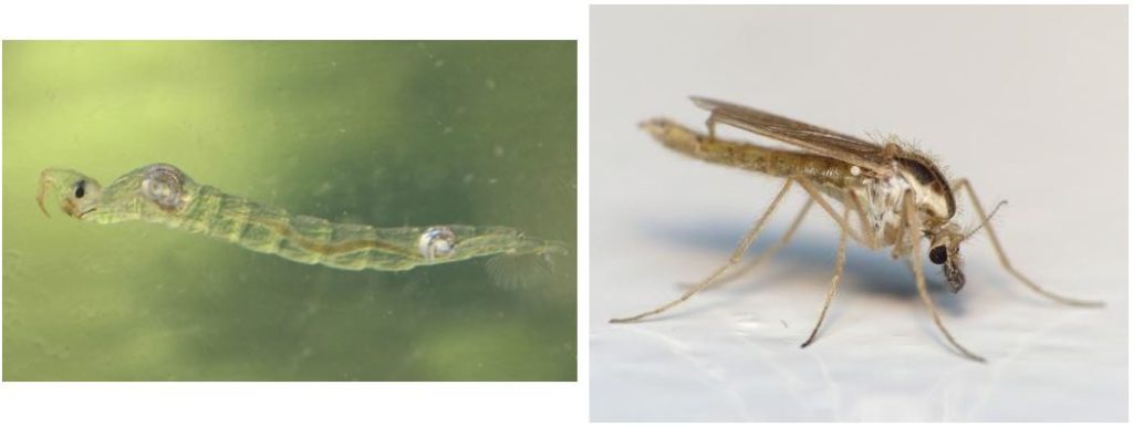
Once the eggs have been deposited near an aquatic habitat, the predatory Chaoborus larvae develop by feeding on small copepodes (small Freshwater crustaceans and seawater), cladoceran (small Freshwater crustaceans where the thorax and abdomen have fused) and unicellular protozoa such as ciliated (presence of vibration eyelashes at their surface). These insects are an essential element of trophic balance by creating predation pressure for a multitude of organisms and a food resource for many species of fish. After 4 successive larval stages, the larvae gain land to metamorphose (pupe stage) and adults emerge for mating and reproducing (pictures 1 and 2).
Chaoborus larvae are studied because of their particular hunting technique. Detected using mechanosensory sensilla, the prey is captured through modified appendages located on the head forming a kind of basket. Although the morphology of these anatomical structures was accurately described, their functioning remained speculative.
A German team led by Sebastian Kruppert provides answers in this article published on March 22, 2019 (link).
To study the predation behaviour of Chaoborus larvae, these scientists filmed the capture of daphnids (small aquatic crustaceans of the cladoceran family) in three dimensions by tomography (imaging technology) and large-scale camera speed in order to reconstruct the kinematic movements of the various appendages involving the capture of the prey.
Description of the cephalic organs
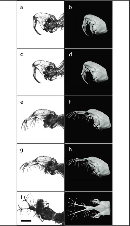
The morphology of the cephalic structures and associated muscles was performed with micro-CT scan (tomography) technology (Figure 1 below).
The head of the larvae forms a sclified capsule of the eucephalous type (an individualised head in an insect larva) consisting of several particularly modified appendages such as mouthpieces and antennae. The mouthpieces are covered on the dorsal side by an elongated rostrum where the antennas are inserted. The labrum, located under the rostrum and the antennas, is equipped with five pairs of bristles. The opening of the mouth, connected to a massive nasopharynx, is surrounded by a pair of Mandibles each with a range of bristles (Photo 3 below).
The antennas are located in the extension of the rostre, the junction grouping also the prelabral appendage. The first antennary segment is as long as the rostre, each of which has four setae (microscopic hairs with various functions: adhesion, camouflage, mechanoreception, communication) curved. The antennas are associated with a contractile muscle. In the resting position, the antennas and setae are steered ventrally, perpendicular to the axis of the rostrum and cover the mouth opening.
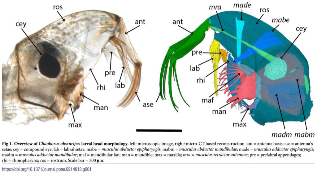
The robust Mandibles are made up of strongly sclified spikes positioned on the ventral and proximal mandibles. These thorns divide into two groups: a dorsal pair with the appearance of bristles, two sturdy and immobile ventral pairs. In the dorsal zone, a range of 10 bristles extends each of the Mandibles (Figure 1 below).
Hunting method
The larvae wait for their prey by floating in the water column. In the resting position, the antennae point ventrally, the Mandibles are closed and the fans folded against the mandibles.
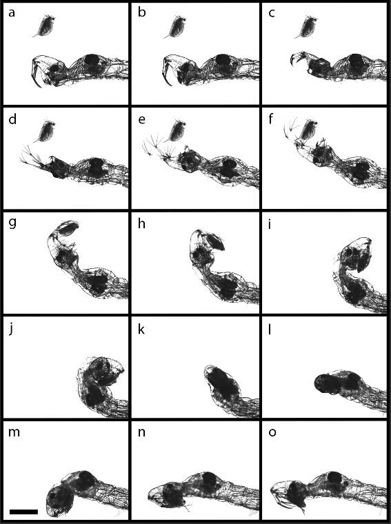
The capture of the prey takes place in four stages:
- Detection of prey by mechanosensory sensilla and compound eyes
- Opening of Mandibles, antennas and setae to form a basket (10.46 MS)
- Capture of the prey by a direction of the basket in its direction then fall back from the basket on the thorax where the fans of the Mandibles prevent the escape of the prey (12.4 MS)
- Contraction of antennae and setae to bring prey to the mouth where it is cheed and swallowed, then return to the starting position (270 MS)
The set of capture steps only lasts 300 Ms (video below).
How structures work
This study also helped to understand how these structures work.
The relief of antennae and setae would likely result from increased hemolymph pressure (same operation as air blast in a long balloon) as no muscle was detected. The return to the resting position would be thanks to the retractile muscle, such as an elastic.
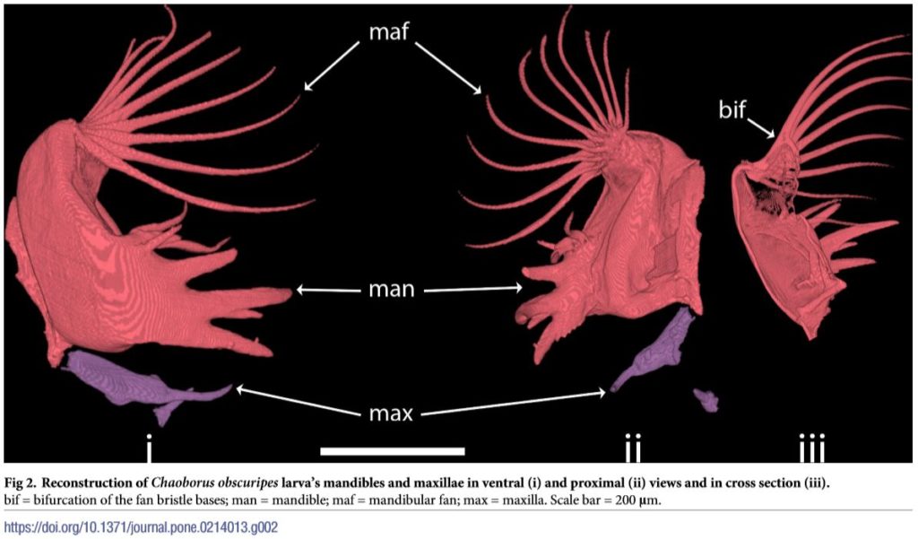
The labrum also tends to increase the pressure of the hemolymph during the capture movement to extend the four pairs of anterior setae and the posterior pair that compose it. Each pair is oriented in a different direction, which closes the catch basket completely. This mechanism is similar for mandible fans (figures 2 and 3).
Conclusion
With a capture movement close to 14 MS, the larva of Chaoborus integrates the Club of the fastest attack movements of the Animal Kingdom.
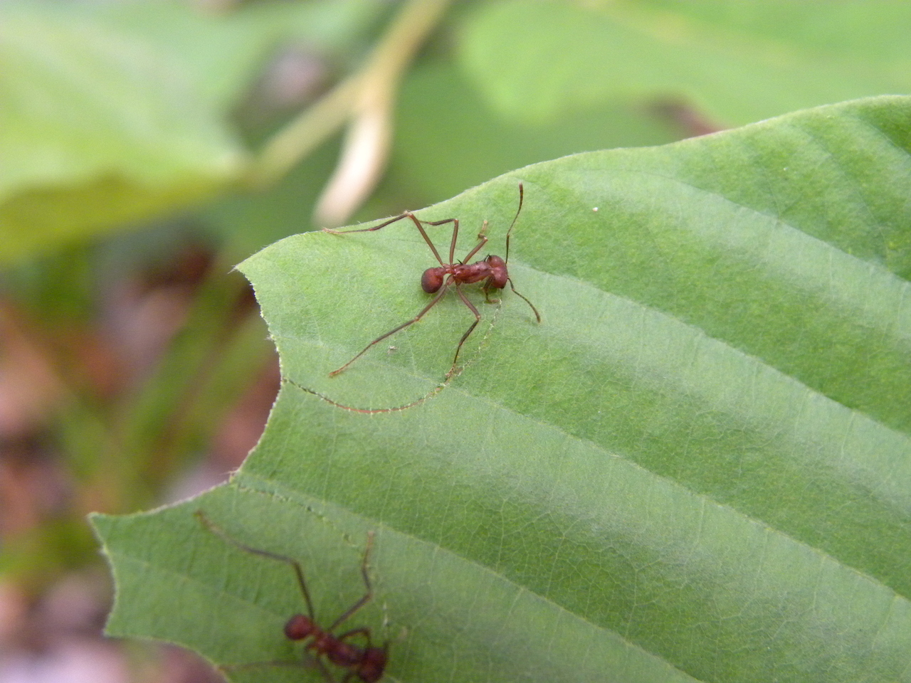
The other members of this Club are the Mantis Coptopteryx viridis (Mantidae) which attacks in 42 MS, the shrimp Squilla empusa (Stomatopoda, Squillidae) and Hemisquilla ensigera (Stomatopoda, Hemisquillidae) attacking in only 4-8 MS as well that the ants of the genus odontomachus (Hymenoptera, Formicidae) who hold the record by triggering an attack in less than 1 DC!
These performances are due to strong selection pressures resulting in co-evolution between prey and predators: evolution pushes prey to become ever faster to escape ever more effective predators.
Source:
- Kruppert S. et al. (2019): Zooplankter’s nightmare: the fast and efficent catching basket of Larval phantom midges-PLOS ONE (link)
Videos
Attack of a Squille
Odontomachus ants
